Scan Identification
- Background
- Contribution of Metadata to Image Signal
- Metadata Informed Scan Identification Process
- Data Dictionaries
- Sequence Dictionary
- Helpful Resources
Background
Scans delivered from CHOP arrive in dicom format. Data is converted to nifti using heudiconv (relies on dcm2niix) where scans were identified(named) based on the SeriesDescription or ProtocolName metadata fields containing explicit mention of various sequence or scan types:
- T1w
- Sequence: tse, spgr, mpr
- Scan: t1w
- T2w
- Scan: t2w
- FLAIR
- Scan: flair
- DWI
- Scan: ep2d, dti
CHOP’s standardized MPR scans would then be identified using CuBIDS and a python script based on these criteria:
MPRStandarized- RepetitionTime = 2.05
- VoxelSizeDim3 = 0.9
- ProtocolName = “T1 MPR SAG 0.9 MM”
MPRStandarizedVariant- RepetitionTime = 2.05
- VoxelSizeDim3 = 0.9
ProtocolName and SeriesDescription are both character strings that can be changed by the scanner operator, leading to an large amount of very small variations (see examples below) and general non-descriptiveness, so it is more reliable to use metadata in the dicoms. Additionally, we want forwards compatability, meaning with each new delivery of data, we want to classify scans with ProtocolName or SeriesDescription that we have never encountered before.
- Some non-descriptive examples
- routine_brain/mts_trans
- routine_brain_1_to_2_years
- ax gre
- sag fspgr
Contribution of Metadata to Image Signal
Our data includes T1w, T2w, FLAIR, PDw, PDT2, DWI, EP2D type images. Image comparison resource
TR and TE
Great resource for understanding TR and TE: link In brief:
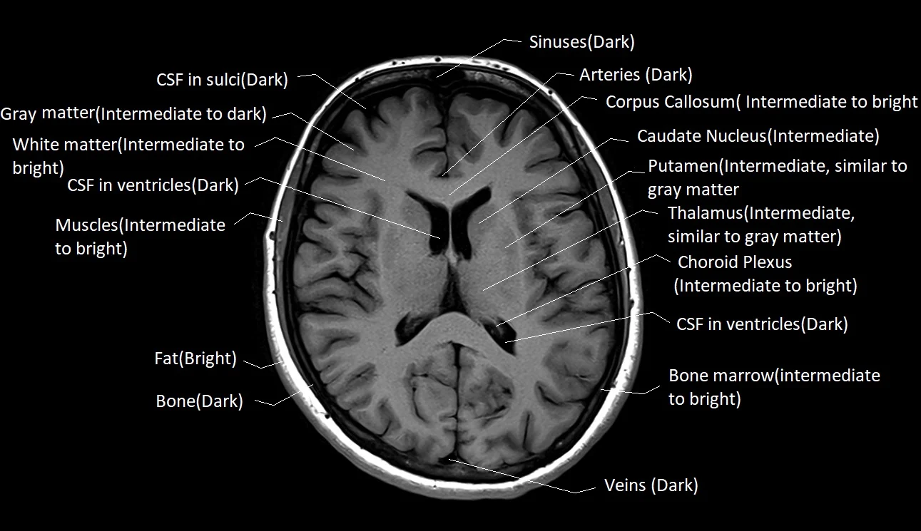
- T1w = short TR and TE
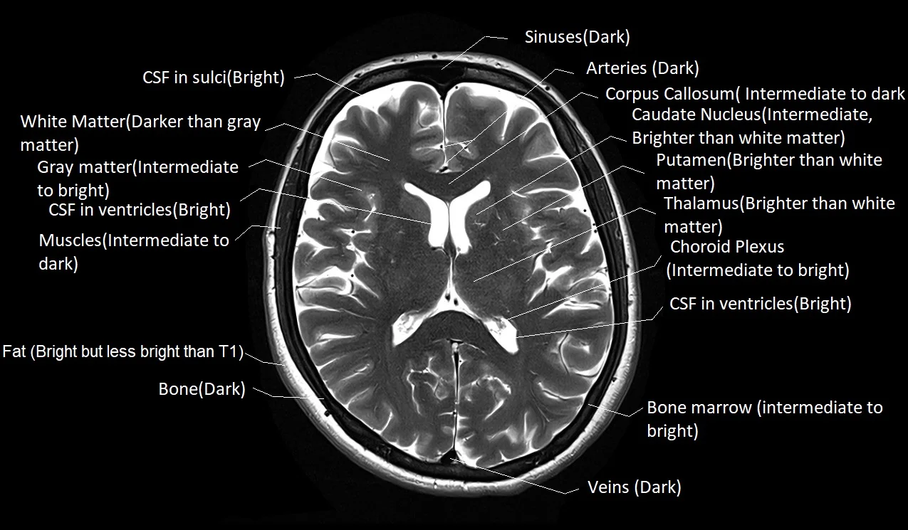
- T2w = longer TR and TE
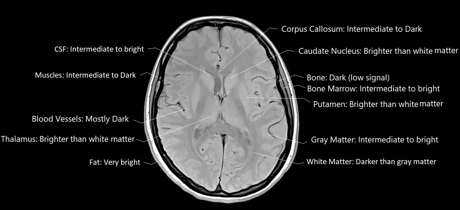
- PD (Proton Density weighted) scans = long TR and short TE
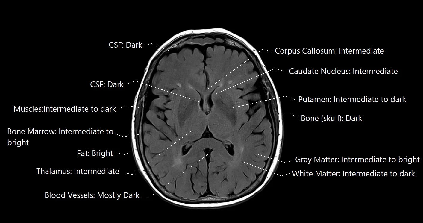
- FLAIR (FLuid Attenuated Inversion Recovery) scans = longest TR and TE
T1w
T2w
PDw
FLAIR
Flip Angle
Explanation of Flip Angle FlipAngle contributes to the brightness of an image. It is a value in degrees between 0 and 180 representative of the angle in which the magnetic field was applied. A higher degree FlipAngle = brighter image, lower degree FlipAngle = darker image
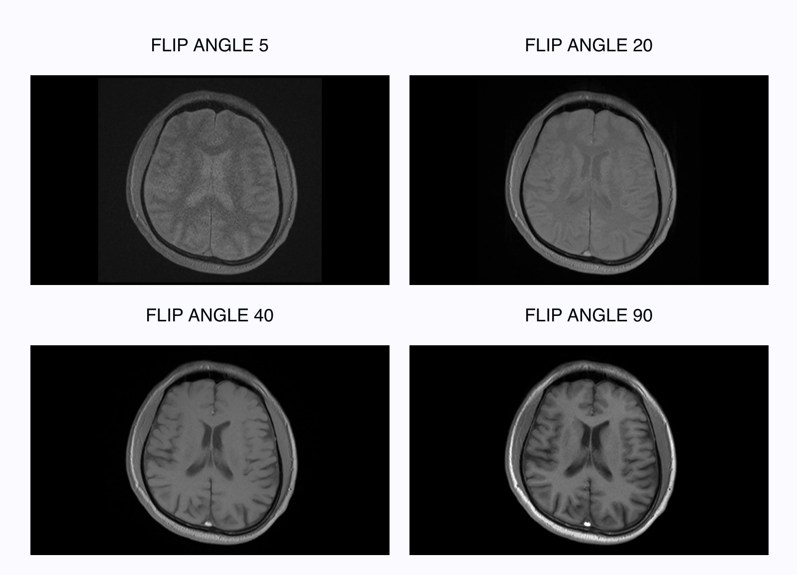
FlipAngle is also associated with Signal-to-Noise Ratio (SNR) and Specific Absorption Rate (as one increases so do the others).
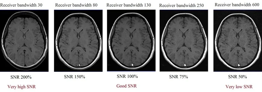
Pixel Bandwidth
Great resource for understanding bandwidth. The PixelBandwidth is equivalent to the Receiver Bandwidth which can be adjusted at the scanner console reflective of the quality of the MR signal. Bandwidth and SNR are inversely proportional, higher bandwidth decreases SNR.
The receiver bandwidth refers to the range of frequencies utilized during the reception of RF pulses.
Percent Sampling
Fraction of acquired acqusition matrix lines.
Multiband Acceleration Factor
Number of slice obtained simultaneously (i.e. value of 4 means 4 slices taken at the same time). Reduces scan time.
Fat Saturation
Under development
Magnetization Transfer
Under development
Metadata Informed Scan Identifcation Process
In order to reliabily classify a scan that might have a non-descriptive or misclassified ProtocolName and/or SeriesDescription, metadata pulled from the dicoms is used to classify a scan into a general scan type (T1w, T2w, FLAIR) and then into a more specific sequence (i.e. TSE T1w, TIRM T2w) based on these fields:
RepetitionTimeEchoTimeFlipAngleImageOrientationPixelBandwidthPercentSamplingMultibandAccelerationFactor
Scans do not always have all of the above fields present due to various archeological reasons such as year of scan (MultibandAccelerationFactor is a result of newer techniques, some older scanners generally output less metadata), scanner manufacturer, etc. At minimum, we expect any scan to have RepetitionTime and EchoTime. The general process for scan identification is described below.
Group Identification
Scan groups were created based on the most common ProtocolNames in the data. These groups can be found in the parameter ranges table. Data was first segmentation by field strength (3T versus 1.5T) and by scan type (T1w, T2w, and FLAIR).
Parameter Review
The metadata fields were chosen in order to distinguish scans belonging to one group from scans belonging to another group. Originally, many metadata fields were considered. These fields were pruned away based on how heterogenous the values were across all scans in that one group. For example, for RepetitionTime, did the majority of scans in that group have the same exact TR? If not, were the majority of TRs a few milliseconds away from the mode (most common value) or were they 4 or 5 second away from the mode? Consider the examples below, the 2 scan groups have very homogenous PixelBandwidth but highly variable RepetitionTime as only 34% and 15% of scans in those groups have a TR within 5 msec of the mode.
| Group | RepetitionTime Mode | % of Scans | PixelBandwidth Mode | % of Scans |
|---|---|---|---|---|
| T2_TSE_COR 384 | 0.099 | 34 | 205 | 99 |
| T2_TSE_TRA 384 | 0.095 | 15 | 205 | 99 |
TR is an essential parameter in distinguishing scan types so even though its highly heterogenous in the examples, it is retained. Other fields that were highly heterogenous were discarded. The figures below will show heterogenity in the retained parameters. In the example it is also shown that while homogenous, PixelBandwidth is the same between the two groups so it isn’t useful for distinguishing these groups apart from each other (mainly because they are both TSE sequences).
Surviving parameters:
RepetitionTimeEchoTimeFlipAngleImageOrientationPercentSamplingPixelBandwidthMultibandAccelerationFactor
Evaluate the heterogenity within each parameter here: Parameter Plots
Classifier Updates
The initial classifer function only identified and labelled CHOP standard MPRAGE with an exact RepetitionTime of 2.05, exact VoxelSizeDim3 of 0.9, and exact ProtocolName of “T1 MPR SAG 0.9 MM”. If scans passed the first two filters and the ProtocolName contained the substring listed previously, it was labelled as a “StandardizedVariant”. To be able to identify scans that resemble one of the groups defined in the previous step, the classifer function had to be updated to allow for filters for all parameters that survived the parameter review step. Additional, a range had to be determined in order to identify scans that very closely matched the pre-defined group parameters (“StandardizedVariant”) and as scans that loosely matched the pre-defined group parameters (“Variant”).
-
Initial filters require an input scan to first match to a group’s field strength (3T or 1.5T)
-
Then:
- “StandardizedVariant” scan matches to a single protocol group based on:
RepetitionTimewithin 0.05 s of the group’s value modeEchoTimewithin 0.01 s of the group’s value modeFlipAngleexactly the first or second most common value for the group, or the parameter doesn’t exist in the scan metadataPercentSamplingexactly the first or second most common value for the group, or the parameter doesn’t exist in the scan metadataPixelBandwidthexactly the first or second most common value for the group, or the parameter doesn’t exist in the scan metadataMultibandAccelerationFactorexactly the first or second most common value for the group, or the parameter doesn’t exist in the scan metadata
- “Variant” scan matches to a single protocol group based on all the same above factors, except:
RepetitionTimewithin 0.5 s of the group’s value modeEchoTimewithin 0.05 s of the group’s value mode
- “StandardizedVariant” scan matches to a single protocol group based on:
Performance Review
Evaluating the performance of the classifer was necessary to ensure that:
- No cross scan type classification (clearly T1w scan passing through filters for a T2w scan group)
- No multi-group classification (no 1 scan being classifed as 2 different groups, i.e. TSE and MPRAGE)
Input data was the same used to define the initial groups.
Example
Consider a scan (done at 1.5T that wasn’t included in the original data to define the groups) with a non-descriptive ProtocolName of ROUTINE_BRAIN_CONTINUED/CO with the following parameters:
RepetitionTime= 6.0EchoTime= 0.097FlipAngle= 180ImageOrientation= LSP+PercentSampling= 100PixelBandwidth= NoneMultibandAccelerationFactor= 30
We could infer that this probably isn’t a T1w or FlAIR, but can’t really know if its T2w or PDw. Putting this scan through the classifer, we see that it matches into a TSE T2w scan group (specifically ROUTINE_BRAIN/COR_TSE_T2 group) with the following definitions:
RepetitionTime= 6.0EchoTime= 0.099FlipAngle= 180ImageOrientation= LSP+PercentSampling= 100PixelBandwidth= NoneMultibandAccelerationFactor= 30
Now we have classifed a previously “unknown” scan using only metadata!
Data Dictionaries
This section will list all of what can show up in clincal data organized after September 2024.
Sample
- BIDS pair:
sample-fetal - Fetal neuro scans
Fat Saturation
Reconstruction Methods
- BIDS pair:
rec-<value> - Values:
- PROPELLER
- reference
- Periodically Rotated Overlapping ParalleL Lines with Enhanced Reconstruction
- NORM
- Intensity normalization
- DIS2D
- Distortion correction in 2D
- DIS3D
- Distortion correction in 3D
- MFSPLIT
- FIL
- In-line filter
- PROPELLER
Sequences
- BIDS pair:
acq-<value> - Values:
- MPRAGE
- Magnetization Prepared Rapid Gradient Echo Imaging
- BLADE
- TSE
- Turbo Spin Echo
- GE
- Gradient Echo
- SE
- Single Echo
- HASTE/SSFSE
- Reference
- HASTE: Half-Fourier Acquisition Single-shot Turbo spin Echo imaging (Siemens)
- SS-FSE: Single-shot fast spin echo (GE)
- SPACE
- CUBE
- VISTA
- VIBE
- TIRMDARK
- Reference
- Turbo Inversion Recovery Magnitude
- TIRMDARKIPAT
- FL2D/SPGR/FLASH/FFE
- Reference
- FL2D/FLASH: 2-dimensional fast low angle shot (Siemens)
- SPGR: spoil gradient recalled echo (GE)
- FFE: T1-fast field echo (Philips)
- This can sometimes show up in the
ImageTypefield
- This can sometimes show up in the
- FL3D
- FL3D/FLASH: 3-dimensional fast low angle shot (Siemens)
- MTS
- Reference
- Magnetization Transfer Saturation
- FSGPRBRAVO
- FSPGR
- TSE3DVFL
- TSEP2
- MEMP
- FSE
- SEPAT2
- PDT2
- TIRM
- STIR
- Reference
- Short Tau Inversion Recovery
- FL3D
- SPC
- CINE
- DIXON
- MPRAGE
Suffix
- Values:
- structural
- T1w
- T2w
- T2starw
- FLAIR
- Fluid Attentuated Inversion Recovery
- T1 v T2 FLAIR
- PDw
- Proton density weighted
- susceptibility
- swi
- diffusion
- dwi
- ep2d
- other
- unknown
- structural
Helpful Resources
General
- What is a nifti?
- Common Abbrevations Dictionary
- What is Inversion Time
- What is SNR
- Magnetization Transfer Contrast
- Cross Vendor Terms
Manufacturer Specific Guides
Siemens
Last updated 2024-10-01 by jmschabdach, originally written 2024-09-27 by dabrielz